Collateral Damage in X-ray Free Electron (XFEL) Experiments
New findings in Nature Communications and Physical Review Research
X-ray free-electron lasers (XFELs) are novel X-ray sources that produce extremely intense yet very brief X-ray pulses, thereby permitting breakthrough experiments and new insights in many fields of science. The pulse duration can be as short as a few femtoseconds, namely only a millionth of a billionth of a second. This allows, for example, molecular reactions to be "filmed" as they happen, yielding unique information about the dynamics of molecular mechanisms and chemical processes. Such information is interesting not just from a basic science point of view but also in facilitating, for example, the design of improved medical drugs, catalysts or batteries.
Performing experiments at XFELs is accordingly of great interest to a large number of scientists worldwide. Unfortunately, access to XFELs is severely limited, since only a handful of these large, kilometre-sized facilities exist around the world. A further bottleneck has been the low pulse generation rate of first-generation XFELs, typically only 30-120 pulses per second, which limits the rate of data collection. Next-generation XFELs based on superconducting technology offer a more than 30,000-fold increase in pulse rate (up to 4.5 million pulses per second, generally denoted as 4.5 MHz). If this increased pulse rate could be fully utilized, it would result in vastly higher data collection rates and correspondingly shorter experiments, allowing greatly increased access to XFEL measurements for prospective users and making many more experiments possible.
Explosive shots

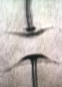
More pulses per second necessarily means a shorter time between the X-ray pulses, 222 nanoseconds at 4.5 MHz. Performing experiments at this tremendous speed naturally presents new experimental challenges. In particular, the X-ray detector and data collection system must be able to handle the veritable deluge of collected X-ray data and the experimental sample must be delivered to the X-ray beam at a commensurate rate. In the case of biological samples, the high-rate sample delivery is performed by means of very fast, microscopically thin liquid jets carrying micron-sized particles of the sample. To investigate such tiny samples a huge X-ray pulse intensity is required, of a strength such that exposure to a single X-ray pulse not only vaporizes the sample particle but also explosively opens a gap in the surrounding jet. The jet explosion can launch a pressure wave travelling along the jet. If two XFEL pulses are spaced too closely in time, the second pulse may interrogate sample that has been altered by the pressure wave generated by the first. Since pressure is a fundamental thermodynamic parameter, an explosion-induced pressure pulse can thereby modify the sample. Even worse, some of the pressure-induced changes (such as damage or deformation) may persist after the pressure pulse. If so, the measured molecular structure and reaction dynamics might correspond to a state different from the native, ambient state in which biological samples function. This problem could seriously affect the way XFEL experiments have to be performed. In particular, the need to avoid high-pressure artefacts could limit the maximum XFEL pulse rate for useful data collection to a fraction of the highest pulse rate available at next-generation XFELs.
The findings
In two papers, recently published in Nature Communications and in Physical Review Research, scientists from the department of Biomolecular Mechanisms at the Max Planck Institute for Medical Research in Heidelberg together with an international research team led by Ilme Schlichting, director at the Max Planck Institute, and by Claudiu Stan, Assistant Professor at Rutgers University-Newark, investigated the effect of explosion-induced pressure waves in a jet on crystals of biological molecules (lysozyme and haemoglobin) carried by the jet. These are two important model systems; their structures are well known under ambient conditions; like all macromolecular crystals they are soft crystals, which makes them vulnerable to damage by the pressure waves. “We showed that the shockwave triggered by the first of two closely spaced X-ray pulses can indeed negatively affect sample integrity,” says Marie Grünbein, first author of the two publications. “The effect is not only a degradation of data that reduces the precision of the molecular structure determination, but more importantly, the molecular structure itself has changed.” This could potentially lead to incorrect 3-dimensional structures of the native molecules and thus to incorrect interpretations of their function and dynamics. “These findings show that careful considerations are required for MHz data collection at new and upcoming X-ray FEL facilities. Our results show that high-pressure effects can occur. However, we also show how to detect such effects and we suggest how the experiments can be adjusted to prevent deterioration. This will help scientists to adjust their experiments to mitigate the pressure effects, and suggests how to design novel techniques (such as using extremely thin jets) that can enable the full potential of next-generation XFELs“, says Marie Grünbein.