
Population Imaging of neuronal activity with genetic calcium sensors
Imaging activity in the cortex
 shows fluorescence changes in the soma in response to single and double action potentials (lower panel).")
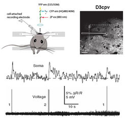
A D3cpv labeled neuron (upper right panel) shows fluorescence changes in the soma in response to single and double action potentials (lower panel).
To image a large number of neurons in layer 2/3 barrel field in vivo, we are using genetic Ca2+ sensor, D3cpv, as a reporter of neuronal activity in mice. With this sensor, we demonstrated that neuronal activity can be detected with single-cell, single-spike resolution (Wallace et al., 2008).
Imaging activity in the olfactory bulb
We generated transgenic mice that show robust expression of fluorescent calcium indicator proteins (FCIPs) in olfactory receptor neurons and granule cells. We then demonstrated that odor-evoked calcium responses can be visualized with high spatial and temporal resolution in these cell types by wide-field imaging in living mice (Hasan et al., 2004).

Figure (a) shows thinned olfactory bulb, (b) single-trial, odor-evoked (2-hexanone) activity map in the olfactory bulb and (c) Ca2+ activity trace (green and red) in two different regions during the time of odor presentation (grey box). Note that Ca2+ activity responses are phase-locked to respiration (black trace).