Biomaterials
The Biomaterials facility provides scientific and technical expertise in microfluidic technology, in particular droplet-based microfluidics for synthetic biology and biomedical applications. Additionally, the facility provides state-of-the-art technologies for in-vitro engineering of human 3-D tissue models that can recapitulate healthy and disease tissue conditions. Our best expertise is in fabrication of scaffold-free skin models and in mimicking several skin ailments ranging from atopic dermatitis, psoriasis and skin wounds. Moreover, the facility provides support with high resolution analysis of biomaterials by means of cryogenic and room temperature scanning electron microscopy. The Biomaterials facility is operating in Stuttgart, and is headed by Dr. Ilia Platzman.
Microfluidics:
Microfluidics combines principles of science and technology, and enables the user to handle, process and manipulate fluids of very small volumes – down to less than a few picoliters – via microchannels. This technology permits the integration of multiple laboratory functions into one single microfabricated chip, requires minimal manual user intervention and sample consumption, and allows for fast data analysis and very high precision. For fundamental and applicable, biomedical, biophysical and synthetic biology research the Biomaterials facility provides the following scientific and technical expertise: design and development of various microfluidic functional units (e.g., droplets generator, observation, sorting and picoinjection); laser lithography; clean room operation; and analysis of the microfluidic devices’ performances.
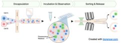
Specifically, we are experts in the capacities of the microfluidic technology technology in the following topics: (1) Bottom-up assembly of bio-inspired synthetic cells (Figure 1) and (2) Mimicry of the cellular environment for personalized cell therapies (figure 2).


Skin tissue models:
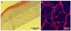
Worldwide, there is a consensus in the need to reduce and replace animal models. Therefore, tissue engineered in-vitro human 3-D models that can recapitulate healthy and disease native tissue are in high demand. Skin is the largest organ of the human body and the primary barrier against the external environment, protecting the internal organs from chemicals, radiation, and pathogens. There are several incurable skin diseases that manifests from environmental and genetic factors. The Biomaterials facility provides expertise in fabrication of scaffold-free skin models and mimicry of several skin ailments ranging from atopic dermatitis, psoriasis and skin wounds. Scaffold-free models are prepared by single cell extracellular matrix (ECM) coating and accumulation technique where human dermal fibroblasts are coated layer-by-layer with ECM moieties and assembled. The assembled cells rapidly form thick underlying dermal compartment. Human epidermal Keratinocytes are then added and the models are cultured in air-liquid interface, thereby mimicking the human skin architecture (Figure 3A). Such scaffold-free skin models can be cultured for long-term with minimal constriction and degradation, which allows testing of novel drugs for longer period of time. Furthermore, the bottom-up approach allows inclusion of other important cell types that reside in native skin. The facility has an expertise to include vascular and immune cells within the skin models (Figure 3B). The fabricated immno-competent and vascularized human skin models can be utilized to mimic several inflammatory skin diseases and as a reliable testing platform for clinically-relevant therapies.
 H&E staining of cryo-sectioned full-thickness human skin model depicting differentiated epidermis with visible cornification B) Confocal image of blood vessels (red, CD31 stained) in the fabricated vascularized skin models (Blue, DAPI stain).")