Population Imaging of neuronal activity with genetic calcium sensors
Imaging activity in the hippocampus
A genetic Ca2+ sensor, GCaMP2, was expressed in rat hippocampal organotypic slices and spatiotemporal population imaging of neuronal activity dynamics in CA3 neurons (left panel) could be detected in single trials (right panel). Simultaneous extracellular electrical recording (lower right panel) of the imaged brain slice is shown in the lower left panel.
In collaboration with Alexandra Von Kameke, Martin Both and Andreas Draguhn in the Department of Physiology at the University of Heidelberg.

Fiberoptic recording of neuronal activity with a genetic Ca2+ sensor in freely-moving mice
To find correlations between cellular signals and animal behavior, a major challenge in experimental systems biology is to perform studies in freely-moving mammals and over an extended period. Among mammals, the mouse is a major genetic model for targeted manipulation of cellular properties. We can demonstrate non-invasive fiberoptic recording of population neuronal activity with a genetic Ca2+ sensor in freely moving mice. Dynamic changes in Ca2+ activity were observed from labeled neurons in the barrel cortex during resting, object-touching and moving, possibly reflecting different behaviorally-related brain states (Murayama M.; Hahn, T.; Kügler, S.; Tang, W.; Sprengel, R.; Nagai, T.; Miyawaki, A.; Larkum, M.E.; Hasan, M.T.: (submitted). The combination of optogenetic and fiberoptic methods appears well-suited for investigating the neuronal basis of animal behaviors, aging and neurological diseases.
Functional expression of YC3.60 in vivo
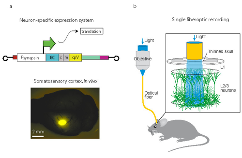
(a) YC3.60 expression in neurons under control of the human synapsin promoter (PhSYN). A fixed mouse showing YC3.60 expression (YFP fluorescence).
(b) Schematic diagram of experimental design. Population Ca2+ activity of neurons in somatosensory cortex were recorded with single core optical fiber.

(c) Fiberoptic Ca2+ recordings from a freely moving mouse. Ca2+ responses (shown in black) and animal behavior (color coded) for an imaging session of 25 seconds are overlayed and specific animal behaviors are indicated on the x-axis with arrow heads.