Resolving the photoswitching mechanism of a fluorescent protein
Combined use of X-ray free-electron laser and spectroscopy provides new insights
Photoswitchable fluorescent proteins are used as molecular markers in super-resolution light microscopy that allows to image living biological cells at a resolution of a few tens of nanometers. These proteins can be reversibly toggled between a non-fluorescent (off) state and a fluorescent (on) state by irradiation with light at specific wavelengths. Photoswitching between on and off states involves ultra-fast excited-state processes that have been recently characterized structurally. Conformational changes on the slower time scale, however, have remained elusive, hampering a comprehensive description of the photoswitching mechanism at the molecular level.
 is excited by a visible-light laser pulse (purple ray) and impacted by an ultra-short X-ray pulse (yellow ray) produced by an XFEL. Ten nanoseconds after excitation of the starting state (off, gray), the protein adopts an intermediate state, the structure of which (cyan) differs from the one of the final state (on, yellow).")
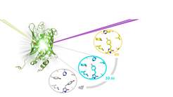
Using time-resolved serial crystallography at the X-ray free-electron laser (XFEL) SACLA in Japan, in combination with transient absorption spectroscopy, researchers from the Department of Biomolecular Mechanisms at the Max-Planck Institute for Medical Research in Heidelberg and collaborators have now established the photoswitching mechanism of rsEGFP2. They determined the three-dimensional structure of a key photo-intermediate which is populated 10 nanoseconds after photoexcitation of the off state (see figure). This study clarifies the order of events during the off-to-on photoswitching and is anticipated to facilitate rational improvement of reversibly photoswitchable fluorescent proteins for applications in super-resolution light microscopy of biological cells.