Introduction to Fiber Diffraction
Fiber diffraction patterns fall into two main classes: crystalline and non-crystalline.

© Kenneth C. Holmes
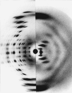
Fig h1: Diffraction from the A and B forms of DNA
© Kenneth C. Holmes
In the crystalline case (e.g. A-form of DNA) The long fibrous molecules pack to form long thin micro-crystals which share a common axis (usually referred to as the c-axis). The micro-crystals are randomly arranged around this axis. The resulting diffraction pattern (on left of figure h1) is equvalent to taking one long crystal and spinning it about its axis during the x-ray exposure. All Bragg reflexions are registered at one time. The reflexions are grouped along "layer-lines" which arise from the repeating structure along the c-axis. However, particularly at high resolution the Bragg reflexions tend to fall on top of each other. If the Bragg reflexions could be separated and measured out to high resolution then the standard methods we have described for crystals could be used. Unfortunately this is never the case and model building must be employed - as is generally the case for non-crystalline fibers.
In non-crystalline fibers (e.g. B-form of DNA) the long fibrous molecules are arranged parallel to each other but each molecule takes on a random orientation around the c-axis. The resulting diffraction pattern (right of figure h1) is also based on layer-lines, which reflect the periodic repeat of the fibrous molecule. The intensity along the layer-lines is continuous and can be calculated via a "Fourier-Bessel Transform" of the repeating structure of the fibrous molecule (the Fourier-Bessel Transform replaces the Fourier transform used in standard crystallography - The Fourier-Bessel transform arises because of the cylindrical symmetry).
