Introduction to Fiber Diffraction
How this applies to DNA

© Kenneth C. Holmes
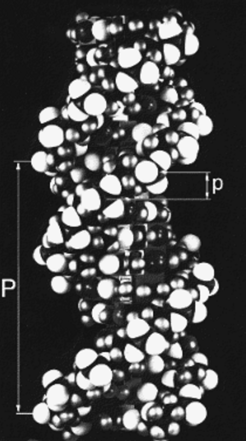
Fig h5: A space filling model of DNA-B form (W. Fuller)
© Kenneth C. Holmes
DNA-B form is a simple helix which repeats in one turn. It has 10 base pairs per turn so that the angle turned ber base Π is 36°. The spacing between the bases is 3.4 Å (i.e. p = 3.4 Å) and the pitch is ten times greater (i.e. P = 34 Å. For low order layer lines the order of the Bessel function n which occurs on the l'th layer line is l. Because of their mass the phosphate groups are the dominant scatterers in a nucleic acids.The phosphate oxygens show up as prominent white spheres in the atomic model (Fig h5). The spacing of the layer lines in Fig h1 corresponds to 34 Å - the helix repeats in 34 Å. In this case this is also the pitch P. Knowing this and Francis Crick's formula for the scattering of a helix, Jim Watson was able to glean the radius of the phosphate groups from the look of the helix cross in Rosalind Franklin's fiber diffraction patterns of DNA . Furthermore, the strong meridional reflexion (see Fig h1) which has a Bragg spacing of 3.4 Å, must correspond to 1/p, (i.e. the spacing between bases was 3.4 Å). These pieces of information went a long way towards defining the essential parameters of the Watson-Crick model.
![DNA-B form is a simple helix which repeats in one turn. It has 10 base pairs per turn so that the angle turned ber base [phi] is 36°. The spacing between the bases is 3.4Å (i.e. p = 3.4 Å) and the pitch is ten times greater (i.e. P = 34Å. For low order layer lines the order of the Bessel function n which occurs on the l'th layer line is l. Because of their mass the phosphate groups are the dominant scatterers in a nucleic acids.The phosphate oxygens show up as prominent white spheres in the atomic model (Fig h5). The spacing of the layer lines in Fig h1 corresponds to 34Å - the helix repeats in 34Å. In this case this is also the pitch P. Knowing this and Francis Crick's formula for the scattering of a helix, Jim Watson was able to glean the radius of the phosphate groups from the look of the helix cross in Rosalind Franklin's fiber diffraction patterns of DNA . Furthermore, the strong meridional reflexion (see Fig h1) which has a Bragg spacing of 3.4Å, must correspond to 1/p, (i.e. the spacing between bases was 3.4Å). These pieces of information went a long way towards defining the essential parameters of the Watson-Crick model.
Fig h5: A space filling model of DNA-B form (W. Fuller)](/34674/original-1310459497.jpg?t=eyJ3aWR0aCI6MzQxLCJmaWxlX2V4dGVuc2lvbiI6ImpwZyIsIm9ial9pZCI6MzQ2NzR9--6a0d8eb97e71e4f902256c2f63cbfd0f1ccae578)